VEGF in AMD history shaped decades of research by prompting a closer look at how growth signals affect the eye. This piece traces the idea from an early 1992 hypothesis to a 1994 discovery, highlighting how histopathology helped bridge guesswork and fact. By looking back, readers can see how basic science and clinical observation grew together to illuminate neovascular AMD.
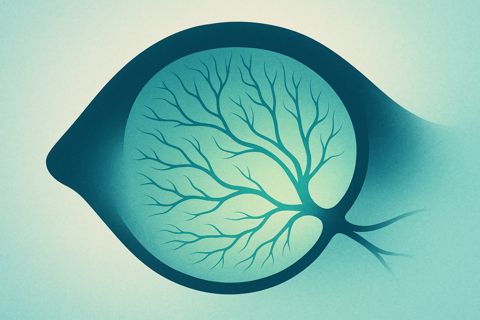
VEGF stands for vascular endothelial growth factor, a signal that encourages blood vessels to sprout where they are not needed. In the eye, this can mean the growth of abnormal, fragile vessels under the retina, a hallmark of neovascular AMD. These vessels can leak plasma and fluid, sometimes causing distortion or loss of central vision. The broader story shows how a molecular signal translates into tissue changes that clinicians monitor and researchers study.
Researchers describe VEGF as a key driver of angiogenesis, the process of new vessel formation. When VEGF activity is high in the macular region, it can create a path for these abnormal vessels to invade the retinal layers. Understanding this mechanism helped scientists connect what they see in tissue with why some patients experience rapid changes in vision.
From 1992 hypothesis to 1994 discovery
The 1992 hypothesis proposed a direct link between VEGF activity and the neovascular changes seen in AMD. It was an idea that could be tested through tissue studies and early experiments, offering a clear target for investigation. By 1994, accumulating observations began to align with the proposal, turning a theoretical concept into a demonstrable finding that researchers could examine in prepared specimens and experimental models. This transition—from a bold guess to evidence-backed insight—helped set the stage for future questions about how to modulate growth signals in the eye.
Histopathology as evidence
Histopathology provided a bridge between the idea and the data. By examining diseased ocular tissue under the microscope, scientists looked for marks of VEGF activity and related vascular changes. The approach offered direct evidence beyond imaging or clinical notes, showing how growth-factor signaling corresponds to the tissue patterns associated with early neovascular AMD. While not the final word, these observations strengthened the case that VEGF plays an active role in the disease process.
Why history matters for today
Understanding this history helps explain why therapies aimed at VEGF and related pathways became central to managing this condition. When researchers trace a path from hypothesis to discovery, they see how a mechanistic insight can move from the lab to the clinic, transforming patient care. The narrative also highlights the value of looking at tissues to interpret signaling pathways, reminding readers that breakthroughs often rest on careful observation and open questions rather than quick answers.
Timeline of key moments
The core arc can be summarized through a couple of moments that are echoed in discussions of the field:
- 1992: A hypothesis proposes VEGF as a driver of AMD-related neovascularization
- 1994: Evidence grows that VEGF is involved, moving the idea toward discovery
Key Takeaways
- VEGF in AMD history marks a shift from guesswork to evidence in understanding neovascular AMD.
- Histopathology provided tissue-level support for VEGF’s involvement in early disease changes.
- The 1994 discovery helped anchor future work on VEGF-targeted therapies.
- Understanding this history underscores how basic science informs clinical strategies in ophthalmology.