Valve orientation is a fundamental part of heart valve surgery. When a prosthetic valve is implanted, correct alignment with the heart’s flow and surrounding tissue matters for how well it works. A scenario where the valve is placed upside down is rare, but it raises important questions about how orientation is checked, what signs could emerge, and how teams respond to ensure patient safety.
Valve Orientation: what it means for the procedure
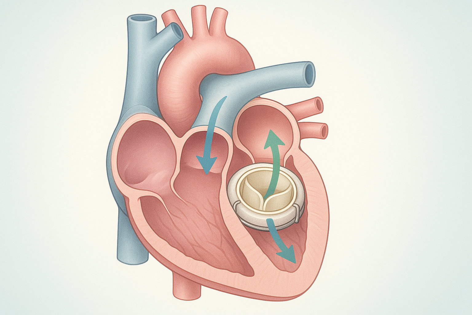
In most valve surgeries, the goal is to position the new valve so that it mirrors normal heart anatomy. The orientation affects how blood moves through the valve and how well the device seals with the heart tissue. If the valve is oriented differently from its intended design, it can change the way it opens and closes. While modern devices and training emphasize correct placement, understanding what orientation means helps patients and families discuss expectations with care teams.
How heart valve implants are placed and checked
Implantation is a carefully planned process that involves imaging, measurements, and intraoperative checks. Surgeons often use imaging guidance and, when appropriate, direct visualization to align the valve with the heart’s anatomy. After placement, tests assess opening, closing, and blood flow through the valve. These checks aim to confirm the valve is oriented and functioning as intended before closing the surgical site.
Recognizing an upside-down implant: possible signs
In rare cases, an implant might be oriented differently than planned. If a valve is not aligned correctly, it can affect how blood flows through the heart or how the device seals with tissue. Some changes may be detected on imaging studies after surgery, while others might be suggested by unexpected murmurs or shifts in heart function. It is important to understand that many orientation issues are identified and corrected during the operation or early in the recovery period with proper imaging and assessment.
Detection, correction, and safety measures
When orientation concerns arise, multidisciplinary teams review intraoperative imaging, device specifics, and the patient’s anatomy. If a problem is detected, surgeons may elect to revise the placement during the same procedure if feasible and safe. Postoperative monitoring, including routine imaging and functional tests, helps confirm that the valve is functioning as intended. Safety protocols emphasize careful verification of orientation and function to reduce the risk of later complications.
Recovery, monitoring, and long-term care
Recovery after valve implantation typically involves a period of monitoring, activity adjustment, and follow-up appointments. Patients may undergo regular imaging to assess valve function and heart performance over time. While the specific plan depends on the valve type and overall health, common themes include ongoing symptom checks, medication decisions tailored to the valve, and lifestyle guidance to support heart health. A patient-centered approach focuses on clear communication about what to expect during healing and how to report concerns promptly.
Key takeaways
- Valve orientation is essential for proper valve function and blood flow.
- Intraoperative checks and imaging help verify correct placement and reduce risks.
- If orientation concerns occur, teams review and may adjust the implant during surgery or early recovery.
- Ongoing follow-up and imaging are important for long-term valve performance and safety.