Understanding volume status is a foundation of many medical decisions. A clear picture of a patient’s fluid status assessment helps explain symptoms, guide testing, and monitor response. This article summarizes what this assessment involves in routine clinical care and why it matters for safe patient management.
What volume status means
In clinical terms, volume status refers to the amount of fluid circulating and available to tissues. Clinicians distinguish states such as dehydration (low fluid volume), hypervolemia (excess fluid), and euvolemia (normal fluid balance). Each state can affect organ function and influence symptoms like dizziness, fatigue, swelling, or shortness of breath. The goal is to describe where a patient sits on this spectrum and how fluid balance may be changing over time.
How clinicians assess fluid status
A systematic assessment blends history, physical findings, and, when available, objective measurements. Clinicians look for signs of fluid loss or gain and for how well the body is maintaining circulation and tissue perfusion.
- History of fluid intake, sweating, vomiting, diarrhea, or poor oral intake
- Changes in urination, thirst, dry mucous membranes, and skin turgor
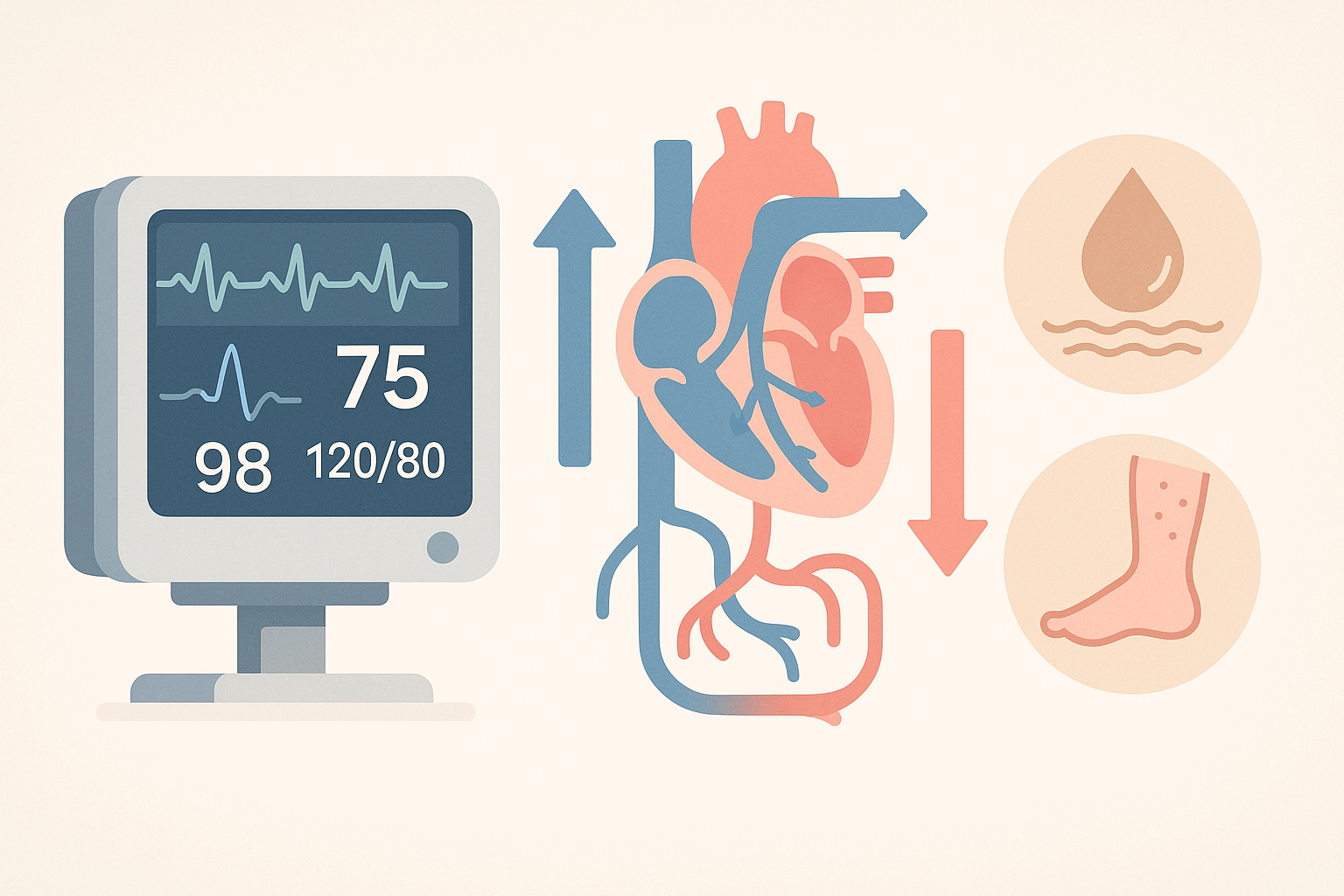
- Vital signs such as heart rate and blood pressure, plus pulse pressure and orthostatic changes
- Physical exam clues like skin elasticity, mucous membrane moistness, edema, and jugular venous pressure when feasible
- Daily body weight and intake/output tracking when available
Interpreting these findings requires context. For example, a high heart rate may accompany dehydration, while edema and crackles in the lungs could point to fluid overload. Clinicians integrate multiple signals rather than relying on a single measure.
Useful tools and tests
Certain bedside tools help refine the assessment. The choice of tests depends on the setting and patient risk.
Bedside ultrasound
Point-of-care ultrasound can provide information about fluid status without invasive testing. One common approach is to assess the diameter and collapsibility of the inferior vena cava (IVC) as an indirect marker of venous return. Results must be interpreted in the clinical context, as several factors can influence ultrasound findings.
Dynamic tests and monitoring
Dynamic approaches, such as a passive leg raise while monitoring hemodynamics, help gauge how the circulation responds to a transient fluid shift. Daily weight, serial vital signs, and careful tracking of urine output add objective context to the clinical picture. In some settings, laboratory clues—such as kidney function tests, serum osmolality, or markers like B-type natriuretic peptide (BNP)—may support the assessment but are not definitive on their own.
These tools are adjuncts to clinical judgment. They help form a picture of volume status without replacing the need for a careful, patient-specific evaluation.
Common patterns and how they differ
Different volume states produce different patterns in signs and symptoms. In dehydration, patients often feel thirsty, have dry mucous membranes, and may show concentrated urine. In dehydration exaggerated by poor intake, orthostatic symptoms and tachycardia may appear. Fluid overload tends to cause swelling in the legs or abdomen, shortness of breath, or elevated pressures on imaging, but even these signs can occur with other conditions. Some patients show a mix of states, which can complicate interpretation. Recognizing the overall trend over time is as important as the snapshot at a single visit.
Monitoring and practical considerations
Ongoing monitoring focuses on changes in signs, symptoms, and objective measures. Clinicians emphasize a patient-specific approach, adjusting assessment methods as the clinical picture evolves. In outpatient settings, simple tools like daily weight and careful intake/output charts can be informative. In inpatient care, continuous monitoring and imaging may be employed to track progress and detect early signs of imbalance.
One key point is that no single test perfectly defines fluid status. The value comes from combining history, examination, and available measurements to create a coherent, evolving assessment. Clear documentation helps healthcare teams track changes and respond appropriately as a patient’s needs shift.
Key takeaways
- Fluid status assessment combines history, exam, and selective tests to describe a patient’s fluid balance.
- Dehydration and fluid overload are common patterns, but many patients have mixed signs that require careful interpretation.
- Bedside tools like ultrasound and dynamic tests can add context but must be interpreted within the broader clinical picture.
- Consistent monitoring (weight, intake/output, vitals) supports timely adjustments and safer care.